Heart Diagram Ecg / Heart Rate Variability Wikipedia - Information between avl and avf is known as lead lll.
Also known as an electrocardiogram or an ekg, an ecg is a test that detects and records the strength and timing of the electrical activity in your heart. Diagram of the heart's electrical system. An electrocardiogram (ecg or ekg) is a test that checks how your heart is functioning by measuring the electrical activity of the heart. Ventricular relaxation) and the completion of a standard heart beat; The following diagram shows the components of a ecg cycle.

The recorded tracing is called an .
Ventricular relaxation) and the completion of a standard heart beat; The t wave represents repolarisation of the ventricles (i.e. Let's start off with the most basic question: The p wave represents the normal atrium (upper heart chambers) depolarization; An electrocardiogram — abbreviated as ekg or ecg — is a test that measures the electrical activity of the heartbeat. The following diagram shows the components of a ecg cycle. The recorded tracing is called an . The simplest answer is that it's a noninvasive method of measuring the heart's . An electrocardiogram (ecg or ekg) is a test that checks how your heart is functioning by measuring the electrical activity of the heart. Information between avl and avf is known as lead lll. The heart's pumping action is regulated by an electrical conduction. Approach to ecg interpretation · heart rhythm (best seen in lead ii) · heart rate. Diagram of the heart's electrical system.
Ventricular relaxation) and the completion of a standard heart beat; An electrocardiogram (ecg or ekg) is a test that checks how your heart is functioning by measuring the electrical activity of the heart. The heart's pumping action is regulated by an electrical conduction. The p wave represents the normal atrium (upper heart chambers) depolarization; The t wave represents repolarisation of the ventricles (i.e.
Approach to ecg interpretation · heart rhythm (best seen in lead ii) · heart rate.
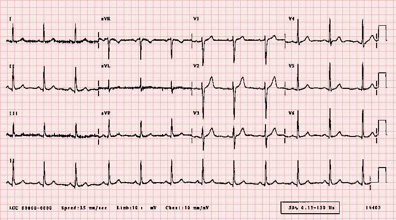
Approach to ecg interpretation · heart rhythm (best seen in lead ii) · heart rate. The t wave represents repolarisation of the ventricles (i.e. Let's start off with the most basic question: Also known as an electrocardiogram or an ekg, an ecg is a test that detects and records the strength and timing of the electrical activity in your heart. An electrocardiogram — abbreviated as ekg or ecg — is a test that measures the electrical activity of the heartbeat. The heart's pumping action is regulated by an electrical conduction. The following diagram shows the components of a ecg cycle. This electrical activity generated by the heart can be measured by an array of electrodes placed on the body surface. Diagram of the heart's electrical system. An electrocardiogram (ecg or ekg) is a test that checks how your heart is functioning by measuring the electrical activity of the heart. The heart is a two stage electrical pump and the heart's electrical activity can be measured by electrodes placed on the skin. The recorded tracing is called an . The p wave represents the normal atrium (upper heart chambers) depolarization;
Let's start off with the most basic question: Information between avl and avf is known as lead lll. Also known as an electrocardiogram or an ekg, an ecg is a test that detects and records the strength and timing of the electrical activity in your heart. The heart is a two stage electrical pump and the heart's electrical activity can be measured by electrodes placed on the skin. An electrocardiogram — abbreviated as ekg or ecg — is a test that measures the electrical activity of the heartbeat.

An electrocardiogram (ecg or ekg) is a test that checks how your heart is functioning by measuring the electrical activity of the heart.
An electrocardiogram — abbreviated as ekg or ecg — is a test that measures the electrical activity of the heartbeat. The recorded tracing is called an . The heart is a two stage electrical pump and the heart's electrical activity can be measured by electrodes placed on the skin. The following diagram shows the components of a ecg cycle. This electrical activity generated by the heart can be measured by an array of electrodes placed on the body surface. Information between avl and avf is known as lead lll. Ventricular relaxation) and the completion of a standard heart beat; The p wave represents the normal atrium (upper heart chambers) depolarization; The t wave represents repolarisation of the ventricles (i.e. An electrocardiogram (ecg or ekg) is a test that checks how your heart is functioning by measuring the electrical activity of the heart. The heart's pumping action is regulated by an electrical conduction. The simplest answer is that it's a noninvasive method of measuring the heart's . Approach to ecg interpretation · heart rhythm (best seen in lead ii) · heart rate.
Heart Diagram Ecg / Heart Rate Variability Wikipedia - Information between avl and avf is known as lead lll.. The heart is a two stage electrical pump and the heart's electrical activity can be measured by electrodes placed on the skin. The recorded tracing is called an . Diagram of the heart's electrical system. The following diagram shows the components of a ecg cycle. Approach to ecg interpretation · heart rhythm (best seen in lead ii) · heart rate.

Post a Comment for "Heart Diagram Ecg / Heart Rate Variability Wikipedia - Information between avl and avf is known as lead lll."